
Navigate through the website by clicking on the icons at the top.
Now that we have mastered incredible GFP technology, our next stop: global domination & world peace!
Fig. 1.2 Section 1.2: History of GFP The timeline below depicts the history and the discovery of GFP, and the brilliant scientists who contributed to its discovery. Click on the link below for a larger and clearer version: Fig 1.3 Click here for a larger and clearer diagram of the timeline. Section 1.3: Structure of GFP Fig 1.5: a scorpion fluorescing under UV light Fig 1.6: a cool animation comparing a genetically-engineered rat glowing with the help of GFP, and a normal wild-type Section 1.5: Incorporation of GFP into Escherichia coli cells 1. Aequorea Victoria cells were broken down to obtain genomic DNA that contain genes encoding for GFP. 7. The E. coli cells that contained gene of interest could be verified by placing under UV light. The arabinose present in the agar plate would activate the GFP gene to fluoresce under UV light.
suffered worked hard for! This section encompasses:  Fig. 1.1
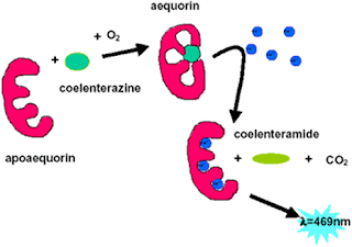
Green fluorescent is a fascinating protein from a species of jellyfish, called Aequorea victoria that has been used in many applications as a tool in the field of biological science. It will result the conversion of blue protein, aequorin, into green fluorescent light by energy transfer. The fluorescence does not come from any chemical cofactor, but from an intrinsic fluorophore created by a cyclization reaction of polypeptide backbone.
The detailed mechanism on how aequorin is converted into green fluorescent light by energy transfer of GFP is shown in the diagram below: 

GFP has a unique can-like shape consisting of an 11-strand β-barrel with a single alpha helical strand containing the fluorophore running through the center. This protein contains 238 amino acids. Below is the beautiful structure of GFP... behold!  Fig. 1.4
With the advance of cloning technology, GFP can be introduced onto many types of living things to serve as an excellent marker for research study and diagnostic purpose. Some of these living things are bacteria, yeast, slime mold, plants, drosophila, zebrafish, and in mammalian cells. It can be used to localize proteins, to study the dynamics of the subcelular compartment to which proteins are targeted. It also reveals new insights regarding physiological activities of living cells.
Here are some animals fluorescing with a bright green glow when illuminated by long-wave ultraviolet light: 


Figure 1.7 shows "Alba" the green fluorescent bunny
2. pGLO plasmid vectors are isolated from E.coli. pGLO plasmid vector contains two genes for selection, one is the ampicillin resistance gene which encodes for B-lactamase and another was the gene encodes for the regulation by the carbohydrate arabinose.
3. The released genomic DNA and pGLO plasmid vectors were then cleaved using the same restriction enzyme.
4. The cut genomic DNA and pGLO plasmid vectors were stitched together by using ligase enzyme.
5. The E. coli cells were being transformed by recombinant vector with gene of interest after ligation step.
6. Only the transformed E.coli cells would then be able to grow on Luria-Bertani agar plate with ampicilin and arabinose due to they contain the genes for selection. Other non-transformed cells would not grow.
8. Once the GFP-producing colonies were isolated, they are innoculated onto another agar plate (with same ingredients) to obtained pure cultures (enrichment method).
Back to Homepage